Hello! Dr. Christina Martini here. This month I want to tell you about an exciting new diagnostic tool we have at SWVH. PennHip helps to diagnose hip dysplasia BEFORE symptoms develop and allows for early intervention to improve the life of your pet!
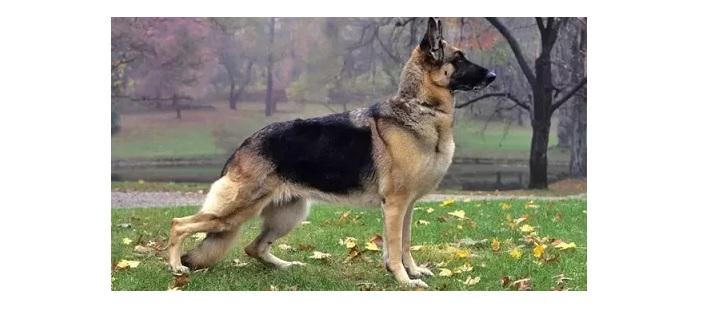
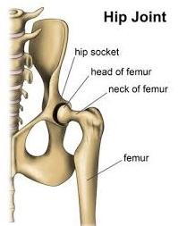
First off, let’s review what hip dysplasia is. Hip dysplasia is a condition where the head or “top” of the femur of the leg does not fit tightly in the acetabulum or “socket” of the pelvis. The creates excessive motion or “laxity” as the head of the femur moves all around the hip socket as your dog walks, runs and plays. This excessive motion creates friction, which over time damages the bone and leads to painful arthritis and disability. Hip dysplasia is a result of both genetics and natural factors. We can try to reduce the amount of hip dysplasia in the population by limiting breeding to only those with good hips.
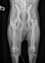
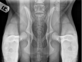
This is where PennHip comes in! PennHip is a special way of taking x-rays to determine the likelihood that your pet will develop hip dysplasia. So, what is actually involved in a PennHip x-ray? First, your pet will receive a complete physical exam. This will include palpation of the hips, testing their range of motion and feeling for any signs of pain or discomfort. Next, your pet is sedated or placed under general anesthesia so they are still and their muscles are relaxed. A total of three x-rays are taken. The first is the traditional ventral/dorsal view of the pelvis with the pet on their back with their hips extended. This is an example of BAD hip dysplasia.
The second image (below) is a view of the patient still on their back but with their hips “frog legged” out to the side and with femurs compressed into the socket.
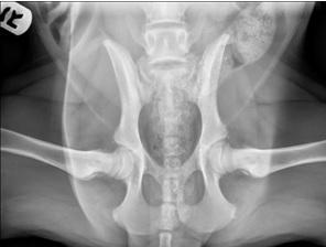
The third and most important view is the “distraction” view created using the PennHip device to open up the space between the head of the femur and the acetabulum of the pelvis (image 3).
Your pet is then woken up and generally does not experience any pain or side effects from the procedure. The x-rays are sent to a board certified veterinary radiologist that is PennHip certified for evaluation and measurements. Specifically, they measure the amount of “laxity” in the hips – the space between the hip socket and head of the femur in the compression versus the distraction view. These measurements are then put in a database of thousands of dogs so that your pet is compared to other pets of the same breed and the risk of developing hip dysplasia is determined.
Pennhip radiographs can be done as early as 16 weeks of age. There is an early intervention procedure called JPS that can be performed up to the age of 20 weeks. This procedure can be combined with a spay or neuter surgery to allow only one needed aesthetic procedure. The pubic symphysis of the pelvis is cauterized so that it stops growing, causing the pelvis to rotate inward and allowing the socket of the pelvis to grip the head of the femur more tightly. This leads to less motion in the hip joint and ultimately less arthritis as your pet ages. After 20 weeks of age (5 months) there are other preventative surgeries that can be performed, but they are more involved and must be performed by a board certified surgeon. If hip dysplasia is diagnosed after one year of age then medical management is instituted to try and delay/minimize the progression of arthritis. This includes keeping your pet at a healthy body weight, moderate consistent exercise, and joint supplements. For severe hip dysplasia, a hip replacement can be considered although this is a very major surgery that only specialty facilities such as UC Davis can perform.
One of the very exciting aspects about PennHip is the ability to try to decrease the prevalence of hip dysplasia in new generations by selective breeding. We recommend PennHip radiographs prior to breeding your dog, and then only breeding if your dog’s hips fall within the top 50% of the breed. That way over time future generations of dogs can lead healthier lives.
You may have heard of the other type of hip screening x-rays called “OFA”. While widely accepted, this has the disadvantage of not being diagnostic until your pet is 2 years of age. This however, is after the window where early intervention surgeries can be performed. In addition, PennHip is much more accurate which is why I consider PennHip to be the superior diagnostic tool for hip dysplasia.
Please feel free to contact us with any questions about this exciting new diagnostic opportunity or to schedule an appointment!